This is the content of the pop-over!


HESI Radiology (HESI-RA) Practice Tests & Test Prep by Exam Edge - Free Test


Our free HESI Radiology (HESI-RA) Practice Test was created by experienced educators who designed them to align with the official Health Education Systems, Inc content guidelines. They were built to accurately mirror the real exam's structure, coverage of topics, difficulty, and types of questions.

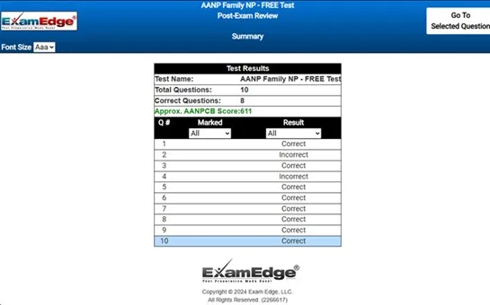
Upon completing your free practice test, it will be instantly reviewed to give you an idea of your score and potential performance on the actual test. Carefully study your feedback to each question to assess whether your responses were correct or incorrect. This is an effective way to highlight your strengths and weaknesses across different content areas, guiding you on where to concentrate your study efforts for improvement on future tests. Our detailed explanations will provide the information you need to enhance your understanding of the exam content and help you build your knowledge base leading you to better test results.

Login or Create an Account to take a free test

After you have completed your free test you will receive a special promo code that will save your between 10-15% on any additional practice tests!


** Sample images, content may not apply to your exam **


Additional test information
Back To General Exam Info

HESI Radiology - Free Test Sample Questions

What is the earliest time that a fetal ultrasound can detect a myelomeningocele?


Correct Answer:
by the 17th or 18th week of gestation


myelomeningocele, a severe form of spina bifida, is a congenital defect characterized by the protrusion of spinal cord tissues, meninges (protective coverings of the spinal cord and brain), and cerebrospinal fluid through an opening in the spine. this condition results from a failure of the spinal canal and backbone to close completely during fetal development, typically within the first month of pregnancy.

the detection of myelomeningocele is crucial for early intervention and management. fetal ultrasonography, commonly known as an ultrasound scan, is a pivotal diagnostic tool used to identify this condition in the womb. by the 17th or 18th week of gestation, detailed anatomical imaging provided by the ultrasound allows healthcare providers to visualize the spine and assess any abnormalities indicative of myelomeningocele. this timing is based on the developmental milestones of the fetus, particularly the formation and closure of the neural tube, which occurs within the first few weeks of pregnancy.

ultrasound is preferred for initial screening due to its safety, non-invasiveness, and accessibility. it provides real-time images that help in assessing the condition of the fetus and can guide further diagnostic procedures if needed. if an ultrasound suggests the presence of myelomeningocele, additional imaging tests such as fetal mri (magnetic resonance imaging) or ct (computed tomography) scans may be recommended. these tests offer more detailed views and can help in planning potential interventions, including surgery.

early detection through ultrasound not only confirms the diagnosis but also aids in the management of the condition, which may include planning for delivery in a facility equipped to handle special needs or even considering fetal surgery options. such interventions aim to minimize the long-term impact of myelomeningocele on the child’s life, emphasizing the importance of timely and accurate prenatal screening and diagnosis.