This is the content of the pop-over!


NCMA RCMAS (RCMAS) Practice Tests & Test Prep by Exam Edge - Free Test


Our free NCMA Registered Clinical Medical Assistant Specialist (RCMAS) Practice Test was created by experienced educators who designed them to align with the official National Certified Medical Assistant content guidelines. They were built to accurately mirror the real exam's structure, coverage of topics, difficulty, and types of questions.

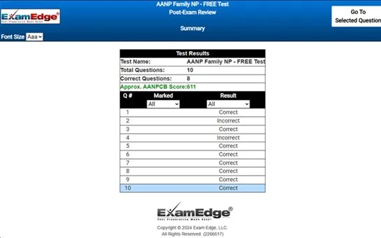
Upon completing your free practice test, it will be instantly reviewed to give you an idea of your score and potential performance on the actual test. Carefully study your feedback to each question to assess whether your responses were correct or incorrect. This is an effective way to highlight your strengths and weaknesses across different content areas, guiding you on where to concentrate your study efforts for improvement on future tests. Our detailed explanations will provide the information you need to enhance your understanding of the exam content and help you build your knowledge base leading you to better test results.

Login or Create an Account to take a free test

After you have completed your free test you will receive a special promo code that will save your between 10-15% on any additional practice tests!


** Sample images, content may not apply to your exam **


Additional test information
Back To General Exam Info

NCMA Registered Clinical Medical Assistant Specialist - Free Test Sample Questions

This test is a radiographic visualization of a joint by injection of contrast media.


Correct Answer:
arthrography


thrography.

arthrography is the radiographic visualization of joints. this diagnostic procedure involves the injection of a contrast medium directly into the joint space to enhance imaging details. the contrast media used are usually iodine-based, which makes the internal structures of the joint, such as cartilage, tendons, and ligaments, visible on x-ray images. this is particularly useful for identifying abnormalities associated with the joint capsule, articular cartilage, and other intra-articular structures like the synovium and joint ligaments. arthrography is commonly used to assess joint conditions such as tears, degeneration, or diseases like arthritis.

in contrast, angiography is the radiographic examination of blood vessels. it involves the injection of a radiopaque contrast agent through a catheter into the bloodstream. the purpose of angiography is to visualize the flow of blood through the arteries and veins to detect abnormalities such as blockages, clots, or malformations.

cholecystography, on the other hand, is a radiographic examination of the gallbladder. for this test, a contrast medium that is absorbed by the liver and excreted into the bile is administered. the concentration of this contrast in the gallbladder allows for detailed imaging of the gallbladder's structure and function, aiding in the diagnosis of conditions like gallstones or cholecystitis.

myelography involves the introduction of contrast media into the subarachnoid space surrounding the spinal cord and nerve roots. this procedure is used to diagnose abnormalities of the spinal canal, including the cord and nerve roots, such as herniated discs, tumors, or stenosis.

each of these procedures—arthrography, angiography, cholecystography, and myelography—utilizes radiographic techniques tailored for visualizing specific body systems to aid in accurate diagnosis and treatment planning. however, for the specific task of visualizing the internal structures of a joint, arthrography is the appropriate procedure.