This is the content of the pop-over!


DHA MRI Technologists (DHA-MRI) Practice Tests & Test Prep - Free Test


Our free DHA Magnetic Resonance Imaging Technologists (DHA-MRI) Practice Test was created by experienced educators who designed them to align with the official Dubai Health Authority content guidelines. They were built to accurately mirror the real exam's structure, coverage of topics, difficulty, and types of questions.

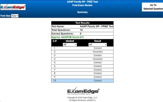
Upon completing your free practice test, it will be instantly reviewed to give you an idea of your score and potential performance on the actual test. Carefully study your feedback to each question to assess whether your responses were correct or incorrect. This is an effective way to highlight your strengths and weaknesses across different content areas, guiding you on where to concentrate your study efforts for improvement on future tests. Our detailed explanations will provide the information you need to enhance your understanding of the exam content and help you build your knowledge base leading you to better test results.

Login or Create an Account to take a free test

After you have completed your free test you will receive a special promo code that will save your between 10-15% on any additional practice tests!


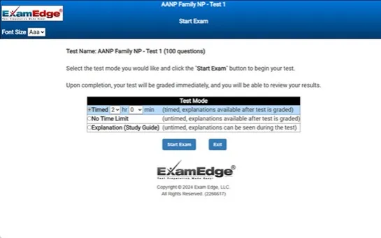
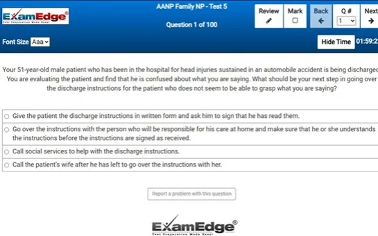
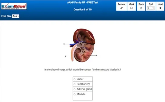
** Sample images, content may not apply to your exam **


Additional test information
Back To General Exam Info

DHA Magnetic Resonance Imaging Technologists - Free Test Sample Questions

A diagnostic imaging technique in which the patient is injected with radioactive glucose, and then images are taken of areas of the body where the high metabolic activity is occurring, is known as which of the following?


Correct Answer:
positron emission tomography.
the correct answer to the question is: positron emission tomography (pet).

positron emission tomography, commonly referred to as pet, is an advanced diagnostic imaging technique used primarily in the fields of neurology, oncology, and cardiology to observe metabolic processes in the body. this technique provides vital information about the physiological functioning of tissues and organs. pet is particularly useful in detecting cancer, monitoring cancer treatment, examining the effects of cardiovascular diseases, and studying brain abnormalities in patients with neurological disorders.

the process begins with the injection of a radiotracer, which typically involves a form of glucose known as fluorodeoxyglucose (fdg), tagged with a radioactive isotope. glucose is a major energy source for most cells in the body, and cells that are most active metabolically (such as cancer cells) will use glucose more rapidly than normal cells. after the injection, the patient waits for approximately one hour to allow the radiotracer to distribute throughout the body and be absorbed by the tissues.

during the pet scan, the patient lies within a donut-shaped scanning machine. this machine detects the gamma rays emitted by the radiotracer. as the radiotracer decays, it emits positrons, which immediately collide with nearby electrons resulting in the emission of gamma rays. these gamma rays are then detected by the pet scanner. the scanner uses this information to create detailed, three-dimensional images of the tissues within the body. areas of high radiotracer uptake appear as bright spots on the pet images, indicating high metabolic activity.

the visualization of these bright spots helps physicians pinpoint areas of abnormal metabolic activity associated with disease. for example, in oncology, pet scans are crucial for detecting cancer metastases, or the spread of cancer to new areas, by identifying rapidly dividing cancer cells that consume glucose at a high rate. in neurology, pet helps in assessing brain functions and diagnosing conditions such as alzheimer's disease by observing cerebral glucose metabolism. in cardiology, pet can assess myocardial perfusion and viability, aiding in decisions regarding revascularization procedures.

overall, pet scans are a valuable tool in the diagnosis and management of various diseases, providing critical information that often cannot be obtained through other imaging techniques. the ability of pet to monitor the effectiveness of treatment also makes it an essential part of modern medical diagnostics, helping to tailor therapies to individual patients' needs and responses.