This is the content of the pop-over!


DHA Vascular Interventional Radiography (DHA-VI) Practice Tests & Test Prep - Free Test


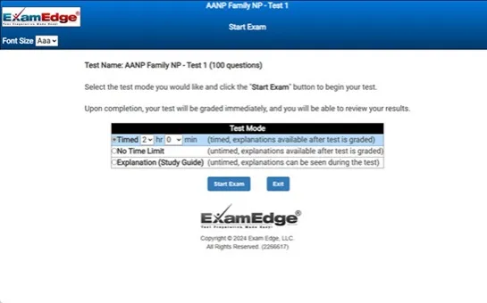
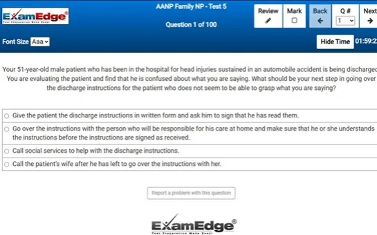
Our free DHA Vascular Interventional Radiography (DHA-VI) Practice Test was created by experienced educators who designed them to align with the official Dubai Health Authority content guidelines. They were built to accurately mirror the real exam's structure, coverage of topics, difficulty, and types of questions.

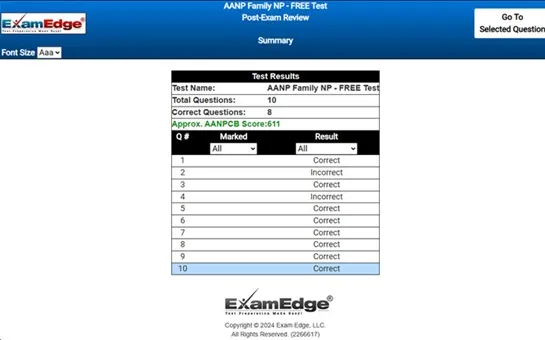
Upon completing your free practice test, it will be instantly reviewed to give you an idea of your score and potential performance on the actual test. Carefully study your feedback to each question to assess whether your responses were correct or incorrect. This is an effective way to highlight your strengths and weaknesses across different content areas, guiding you on where to concentrate your study efforts for improvement on future tests. Our detailed explanations will provide the information you need to enhance your understanding of the exam content and help you build your knowledge base leading you to better test results.

Login or Create an Account to take a free test

After you have completed your free test you will receive a special promo code that will save your between 10-15% on any additional practice tests!


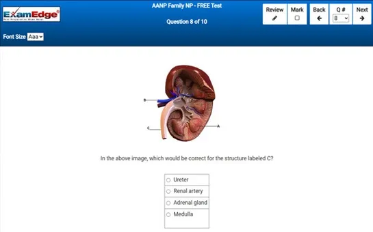
** Sample images, content may not apply to your exam **


Additional test information
Back To General Exam Info

DHA Vascular Interventional Radiography - Free Test Sample Questions

Positioning of the patient for renal venography is done in the:


Correct Answer:

supine position

a renal venograph is an x-ray that is used to evaluate the kidneys. the test can also determine the amount of hormone levels the kidney is producing. further, renal venography is indicated for evaluation of the kidneys for tumors or renal cell carcinoma, blood clots (renal thrombus), for instance. access for catheter insertion is done through the femoral or jugular vein.

positioning of the patient involves placing the patient in the supine position on the x-ray table. the patient's abdomen is centered so that it is over the film. the skin located over the femoral vein, if used for access, is prepped by cleaning the skin using antiseptic liquid and then draped. an anesthetic agent is then injected, followed by cannulation of the femoral vein. fluoroscopic guidance is used where a guide wire is threaded into the vein and a catheter is inserted over the wire and passed into the inferior vena cava.

a test bolus is used to allow view of the vein and to make sure the contrast agent was administered into the vena cava and to check for patency of the catheter. once the catheter patency is checked, the catheter is progressed into the vein where contrast is administered.